
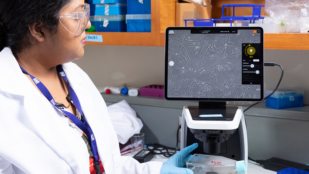
Jhinuk Saha, Ph.D., observes astrocytes under a microscope in a lab at the National High Magnetic Field Laboratory in Tallahassee, Florida. Astrocytes are key glial cells that regulate brain homeostasis and lipid metabolism. Astrocytic lipid dysregulation and reactive states contribute to neurodegenerative pathways, where toxic amyloid‑β interactions disrupt cellular function, drive neuroinflammation, and accelerate neuronal damage in Alzheimer’s disease. (Scott Holstein/FAMU-FSU College of Engineering)
Researchers at the FAMU-FSU College of Engineering and the National High Magnetic Field Laboratory have shown that higher magnetic fields can improve the analysis of molecules linked to Alzheimer’s disease, a finding that could aid the development of future treatments.
Key Points
- A team led by Ayyalusamy Ramamoorthy, a professor at Florida State University in the FAMU-FSU College of Engineering’s Department of Chemical and Biomedical Engineering, published a study in the journal Solid State Nuclear Magnetic Resonance showing that high-field NMR spectroscopy improves the accuracy of measurements of amyloid beta proteins, which are linked to Alzheimer’s disease.
- The team, which included FSU postdoctoral researcher Jhinuk Saha and University of Wisconsin researcher Thirupathi Ravula, analyzed amyloid beta mixed with lipids to more closely mimic conditions inside the human brain than standard lab samples allow.
- The research, announced June 30, 2026, could help scientists design compounds that block the toxic protein interactions driving Alzheimer’s disease, which affects an estimated 7.4 million Americans age 65 and older, according to the Alzheimer’s Association.
- The team plans to continue this work using the National High Magnetic Field Laboratory’s 1.5-gigahertz NMR spectrometer.
In a study published in Solid State Nuclear Magnetic Resonance, the researchers showed that a high-magnitude magnetic field can improve the accuracy of measurements of the chemical composition of amyloid beta fragments, small protein fragments shown to play a critical role in Alzheimer’s disease. They were able to analyze amyloid proteins even when structurally complex and mixed with lipids, creating conditions that more closely resemble those of the human brain than those in traditional laboratory samples.
By better understanding the composition and structure of these molecules, scientists can design compounds that may disrupt disease progression and lead to more effective treatments.
“The current treatment plans for Alzheimer’s disease are not working well enough,” said study co-author Ayyalusamy Ramamoorthy, a professor at Florida State University and in the joint college’s Department of Chemical and Biomedical Engineering. “This disease follows a complex process. We are looking into the mess of molecules implicated in memory loss, investigating how they promote toxic compounds in the brain and trying to stop them.”
How Could This Discovery Help Treat Alzheimer’s Disease?

Researchers are still studying the exact mechanisms that cause Alzheimer’s disease, but amyloid beta proteins are believed to play a central role. These proteins are found clumped together among neurons inside affected brains. Studies have shown them to be a good benchmark for tracking disease progression and a potential target for treatment.
By mapping the structure of amyloid beta catalyzed by lipids, researchers can develop compounds that could effectively bind to its surface and stop it from killing neuronal cells within the brain.
“It’s like an incredibly complex puzzle piece,” Ramamoorthy said. “We want to create another puzzle piece that can match with it and stop it from binding with something within the brain responsible for memory.”
What Did the FAMU-FSU Research Team Discover About Amyloid Beta Proteins?
To find the edges of that puzzle piece, Ramamoorthy and the research team used a nuclear magnetic resonance (NMR) spectrometer. NMR spectrometers work by placing a sample in a strong magnetic field and applying radio waves to excite atomic nuclei. By measuring how atomic nuclei absorb and re-emit these radio waves, scientists can determine properties such as the chemical composition of molecules.
Instead of clean samples, researchers analyzed amyloid beta interacting with a lipid found in the membrane of neural cells. That emulated the tangled mix of cells found within the brain.
They measured samples with a 600-megahertz spectrometer and a 1,100-megahertz spectrometer and compared the results. Researchers already knew that a higher magnetic field would enhance the spectral resolution of amyloid beta proteins. This study showed that an NMR spectrometer operating at a higher magnetic field could also better identify discrete regions of amyloid beta within a realistic sample.
Even though the protein-lipid mix looks chaotic overall, the improved measurements revealed distinct, well-ordered segments within the combined samples and evidence of a central core inside amyloid proteins.
“When you have these amorphous collections of different cell types, they are not well-ordered. When you try to take a picture, it looks very blurry,” Ramamoorthy said. “We were able to zoom in and get a look at the structured regions within the protein.”
Why Does This Alzheimer’s Research Matter?

The study shows that a higher magnetic field NMR spectrometer can identify information from amyloid proteins that exist in a diverse mixture of cell types. Scientists studying Alzheimer’s disease are no longer limited to ideal samples. They can study complex mixtures and still get atomic-level clues.
The researchers plan to use the National High Magnetic Field Laboratory’s 1.5-gigahertz NMR spectrometer for future research.
“This is the only place in the world where such an ultra-high magnetic field (1.5-GHz) NMR spectrometer is available,” Ramamoorthy said. “We want to push the challenges and overcome the hurdles in developing potential drugs to treat Alzheimer’s and related diseases, and these resources are crucial for this work.”
FSU postdoctoral researcher Jhinuk Saha and University of Wisconsin researcher Thirupathi Ravula were co-authors on this study. This research was supported by the National Institutes of Health’s National Institute of Diabetes and Digestive and Kidney Diseases (NIDDK), the National Science Foundation and Florida State University. The research used the National High Magnetic Field Laboratory at FSU and the National Magnetic Resonance Facility at the University of Wisconsin.
Frequently Asked Questions
What is amyloid beta and why is it linked to Alzheimer’s disease? Amyloid beta is a small protein fragment that clumps together among neurons in the brains of people with Alzheimer’s disease. Researchers use it as a benchmark for tracking disease progression and as a potential target for treatment, though the exact mechanisms that cause Alzheimer’s are still being studied.
What is NMR spectroscopy and how is it used in Alzheimer’s research? Nuclear magnetic resonance (NMR) spectroscopy places a sample in a strong magnetic field and applies radio waves to excite atomic nuclei. By measuring how the nuclei absorb and re-emit those radio waves, scientists can determine a molecule’s chemical composition and structure, information that helps them understand how amyloid beta proteins behave in the brain.
What did FAMU-FSU College of Engineering researchers find in this study? A team led by Professor Ayyalusamy Ramamoorthy found that a higher-field NMR spectrometer (1,100 megahertz, compared with a standard 600-megahertz instrument) could identify well-ordered, structured segments and a central core within amyloid beta proteins, even when those proteins were mixed with lipids in conditions that mimic the human brain.
Where was this research conducted? The research was conducted at the National High Magnetic Field Laboratory on the Florida State University campus in Tallahassee, Florida, in collaboration with the FAMU-FSU College of Engineering and the University of Wisconsin’s National Magnetic Resonance Facility.
Who funded this research? The study was supported by the National Institutes of Health’s National Institute of Diabetes and Digestive and Kidney Diseases (NIDDK), the National Science Foundation and Florida State University.
What’s next for this research? The research team plans to use the National High Magnetic Field Laboratory’s 1.5-gigahertz NMR spectrometer, which Ramamoorthy says is the only instrument of its kind available anywhere in the world, to continue studying compounds that could disrupt Alzheimer’s disease progression.
Editor’s Note: This article was edited with a custom prompt for Claude Sonnet 4.6, an AI assistant created by Anthropic. The AI optimized the article for SEO/GEO discoverability, improved clarity, structure and readability while preserving the original reporting and factual content. All information and viewpoints remain those of the author and publication. This article was edited and fact-checked by college staff before being published. This disclosure is part of our commitment to transparency in our editorial process. Last edited: 07/01/2026.
RELATED ARTICLES
Professor to Showcase Groundbreaking NMR and Biomedical Research in France
Research Shows How Insulin, Zinc and Ph Can Block Harmful Protein Clumps Linked to Type 2 Diabetes
FAMU-FSU Engineering Joins Mayo Clinic to Research Alzheimer’s Disease Treatments